Article | May 4, 2025
Tissue AI works like GPS navigation—with real-time traffic data, street-level details, and insights into how people move and interact. It reveals not just where cancer cells are, but how they communicate, evolve, and shape their surroundings—guiding researchers toward more precise and effective treatments.
The human body doesn’t function one cell at a time—it operates as a complex interconnected network of tissues where different types of cells interact in ways that can dramatically influence health and disease. Tissue AI applies artificial intelligence to map and understand what is happening in those tissues, helping researchers uncover new ways to treat disease.
“It's much easier to study a single cell at a time, but that’s not really how the body works. You have all of these cells working together. And the problem is that that’s a lot of information all at once,” says Jeff Chuang, JAX professor and deputy director of the JAX Cancer Center. “It’s why it’s such an exciting time for computational biologists, like myself, to use AI approaches to analyze all this data.”
The human genome has roughly 20,000 genes all giving orders to their cells. Whether those genes are “turned on” and broadcasting instructions to the cell, or “turned off” and silent, impacts how cells behave. With Tissue AI, the researchers see and analyze all of that information in every single cell in their sample—not only the expression of the genes in every cell, but also where those cells are located within the tissue.
We can profile tissues in ways that are really leaps and bounds beyond what we could do in the past," Chuang says. "We can now measure the genes in each cell and their behavior across the entire scope of a tissue. That information can tell us so much about how tumors are behaving and could potentially be treated."
Transcript
Why AI?
According to Chuang, scientists still don’t fully know which tissue structures are the most important for disease outcomes.
“We’re still learning and we want systems that can help us figure out when something is good or bad,” says Chuang.
He likens this capability to facial recognition software—just as security cameras can recognize faces based on learned patterns, AI algorithms can identify abnormal tissue structures that might not be immediately apparent to human eyes. It helps researchers uncover hidden patterns within tissues, offering a more complete picture of how diseases develop and how they might be treated more effectively.
"Pathologists have been looking at spatial data for decades with H&E images of tissues, but the recent technological advancements in data generation in the last five to 10 years have really opened up new areas of biology—first in cells in isolation with advances in single cell RNA-seq and then more recently, single cell spatially-resolved data," says Paul Robson, JAX professor and former director of the JAX Single Cell Biology Lab.
"Tissue AI becomes hugely beneficial because the human eye cannot easily see or interpret such massive amounts of data, let alone find patterns in it," he adds.
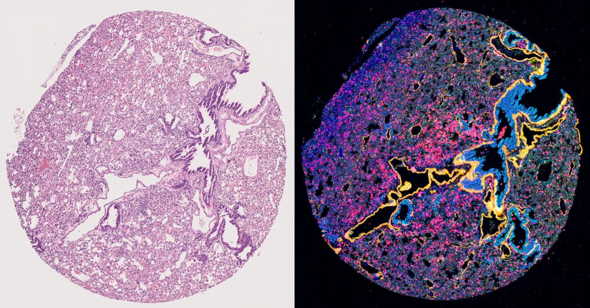
From H&E to AI: Data in technicolor
For more than a century, pathologists have utilized hematoxylin and eosin (H&E) histological slides to diagnose cancer. They take a small biopsy or cutting of a tumor, the sample on a microscopic slide, stain it, and look for the telltale signs of cancer—irregular cell division, and abnormalities in the cell nuclei. They're still considered the gold standard in cancer diagnosis.
Scientists also use bulk assays, which profile DNA from surgically-removed tumor masses and report out average characteristics. Bulk assays are an efficient way to analyze the genetic and molecular structure of a tumor by examining a combined signal from a large population of cells, but they don’t identify the activity of cells in their locations.
“Understanding location-dependence is critical to improve therapies that engage the immune system to kill cancer,” says Chuang.
Tissue AI changes the game by incorporating spatial ‘omics and single-cell imaging—mapping tissue ecosystems in unprecedented detail.
“We provide the tools for researchers to understand biology at the single cell level. And this has been transforming because it has changed completely the way we look at disease, and especially in the context of cancer,” says Elise Courtois, senior research scientist and director of the JAX Single Cell Biology Lab.
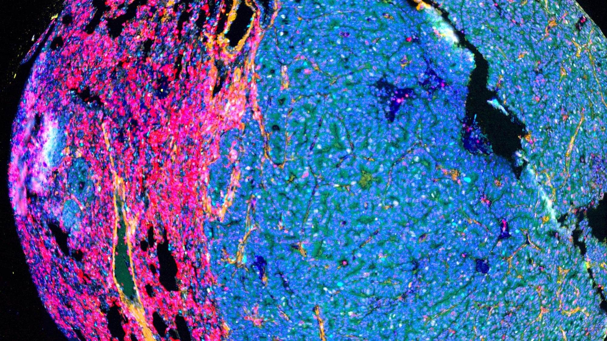
Thanks to the infusion of new data, traditional purple-and-white H&E histological images can be replaced by Tissue AI-generated maps that display thousands of distinct colors—each representing different information.
“Instead of just two colors, we can see 5,000 colors at a time. It is incredibly exciting how much more information we have," Chuang says.
“The past few years have been mind-blowing in terms of technological advancement, allowing researchers to delve deeper into tissue architecture and cellular dynamics with an unprecedented precision. These advances are transforming our ability to map cellular composition, cell phenotyping and function and in ways that were unimaginable just a few years ago,” says Courtois.
Connecting the mouse-human bridge
JAX is part of the Cellular Senescence Network (SenNet), a National Institutes of Health-funded initiative to identify and characterize senescent cells—cells that have stopped multiplying but don't die off when they should. JAX researchers, including Chuang are leading two major SenNet projects, one focused on human tissues and another on mouse models, studying how senescent cells behave in the pancreas, kidney, and placenta.“This is going to create the most awesome cross-species comparison data because it’s the most comprehensive view of the biology spatially-resolved in two different species,” says Robson.“These are ideal platforms to push the human mouse bridge, as well as data sciences, because we're generating huge amounts of data. Presumedly, Tissue AI could help us interpret features in common, as well as unique to either species, and help bridge this human-mouse matrix,” he adds.
Like many researchers in the JAX Cancer Center, Chuang and his team are also focused on the link between cancer and aging. Working alongside JAX scientists Karolina Palucka and Ron Korstanje, they are studying mice aged two years or older, the equivalent of an elderly human. They have been profiling the cancers that appear in those mice to understand how those cancers develop as a function of age. To adapt this research for patients, his team is comparing cancer tissues between mice and people, including projects for both lung cancer and breast cancer.
“We are really at this forefront in terms of being able to compare what's happening in the mouse and human together. It’s something basically no other institute in the world can do the way we can because of JAX’s special strengths in mouse,” says Chuang.
Tissue AI is helping scientists see the full picture, offering a clearer road to better treatments.
This story was featured in Search magazine, Vol. 18, No. 1, published in spring 2025. Search is a publication from The Jackson Laboratory focused on research discoveries and human health.
Learn more
Forum for Discovery
Step into the future of science at JAX’s annual Forum for Discovery on Wednesday, July 9, 2025, where we showcase our groundbreaking research and bold ideas to tackle today’s biggest health challenges and shape a healthier tomorrow.